Histology of Nervous Tissue Review Sheet Answer Key
The Nervous System and Nervous Tissue
Nervous Tissue
Learning Objectives
By the terminate of this section, you volition be able to:
- Depict the basic structure of a neuron
- Identify the different types of neurons on the ground of polarity
- Listing the glial cells of the CNS and depict their role
- List the glial cells of the PNS and describe their office
Nervous tissue is composed of ii types of cells, neurons and glial cells. Neurons are the master blazon of cell that virtually anyone associates with the nervous organization. They are responsible for the ciphering and advice that the nervous system provides. They are electrically active and release chemic signals to target cells. Glial cells, or glia, are known to play a supporting role for nervous tissue. Ongoing enquiry pursues an expanded role that glial cells might play in signaling, but neurons are yet considered the basis of this role. Neurons are important, but without glial back up they would not be able to perform their function.
Neurons
Neurons are the cells considered to be the basis of nervous tissue. They are responsible for the electrical signals that communicate information most sensations, and that produce movements in response to those stimuli, along with inducing idea processes inside the brain. An of import part of the role of neurons is in their structure, or shape. The three-dimensional shape of these cells makes the immense numbers of connections within the nervous system possible.
Parts of a Neuron
Equally you learned in the first section, the main part of a neuron is the cell body, which is also known as the soma (soma = "body"). The jail cell body contains the nucleus and nigh of the major organelles. But what makes neurons special is that they have many extensions of their cell membranes, which are by and large referred to as processes. Neurons are ordinarily described as having one, and only one, axon—a fiber that emerges from the cell body and projects to target cells. That single axon can branch repeatedly to communicate with many target cells. It is the axon that propagates the nerve impulse, which is communicated to one or more cells. The other processes of the neuron are dendrites, which receive information from other neurons at specialized areas of contact called synapses. The dendrites are usually highly branched processes, providing locations for other neurons to communicate with the jail cell trunk. Data flows through a neuron from the dendrites, beyond the cell body, and downward the axon. This gives the neuron a polarity—pregnant that data flows in this one direction. (Figure) shows the relationship of these parts to one another.
Parts of a Neuron
The major parts of the neuron are labeled on a multipolar neuron from the CNS.
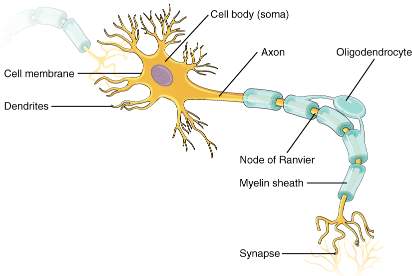
Where the axon emerges from the prison cell torso, there is a special region referred to as the axon hillock. This is a tapering of the cell body toward the axon cobweb. Inside the axon hillock, the cytoplasm changes to a solution of express components called axoplasm. Because the axon hillock represents the beginning of the axon, it is also referred to as the initial segment.
Many axons are wrapped by an insulating substance called myelin, which is really made from glial cells. Myelin acts as insulation much like the plastic or safety that is used to insulate electrical wires. A cardinal difference between myelin and the insulation on a wire is that there are gaps in the myelin covering of an axon. Each gap is called a node of Ranvier and is of import to the way that electric signals travel downwardly the axon. The length of the axon between each gap, which is wrapped in myelin, is referred to as an axon segment. At the end of the axon is the axon terminal, where there are usually several branches extending toward the target prison cell, each of which ends in an enlargement called a synaptic end bulb. These bulbs are what make the connexion with the target cell at the synapse.
Visit this site to learn virtually how nervous tissue is composed of neurons and glial cells. Neurons are dynamic cells with the ability to make a vast number of connections, to respond incredibly quickly to stimuli, and to initiate movements on the basis of those stimuli. They are the focus of intense inquiry considering failures in physiology can lead to devastating illnesses. Why are neurons only found in animals? Based on what this article says about neuron function, why wouldn't they be helpful for plants or microorganisms?
Types of Neurons
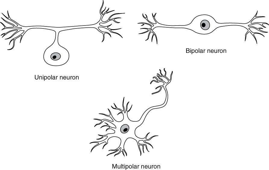
There are many neurons in the nervous arrangement—a number in the trillions. And there are many different types of neurons. They can be classified by many different criteria. The outset manner to classify them is by the number of processes attached to the cell body. Using the standard model of neurons, one of these processes is the axon, and the residual are dendrites. Because information flows through the neuron from dendrites or prison cell bodies toward the axon, these names are based on the neuron's polarity ((Figure)).
Neuron Classification by Shape
Unipolar cells have one procedure that includes both the axon and dendrite. Bipolar cells take ii processes, the axon and a dendrite. Multipolar cells have more than two processes, the axon and ii or more dendrites.
Unipolar cells have only ane process emerging from the cell. True unipolar cells are merely constitute in invertebrate animals, so the unipolar cells in humans are more accordingly called "pseudo-unipolar" cells. Invertebrate unipolar cells do not have dendrites. Human unipolar cells have an axon that emerges from the jail cell torso, but information technology splits so that the axon tin can extend along a very long distance. At one end of the axon are dendrites, and at the other end, the axon forms synaptic connections with a target. Unipolar cells are exclusively sensory neurons and accept ii unique characteristics. Showtime, their dendrites are receiving sensory information, sometimes directly from the stimulus itself. Secondly, the prison cell bodies of unipolar neurons are e'er found in ganglia. Sensory reception is a peripheral function (those dendrites are in the periphery, perchance in the pare) so the cell torso is in the periphery, though closer to the CNS in a ganglion. The axon projects from the dendrite endings, past the prison cell body in a ganglion, and into the fundamental nervous system.
Bipolar cells have ii processes, which extend from each end of the cell body, contrary to each other. One is the axon and i the dendrite. Bipolar cells are not very mutual. They are institute mainly in the olfactory epithelium (where smell stimuli are sensed), and as part of the retina.
Multipolar neurons are all of the neurons that are not unipolar or bipolar. They have i axon and two or more dendrites (usually many more). With the exception of the unipolar sensory ganglion cells, and the 2 specific bipolar cells mentioned above, all other neurons are multipolar. Some cutting edge research suggests that sure neurons in the CNS do non adapt to the standard model of "one, and just one" axon. Some sources describe a quaternary type of neuron, chosen an anaxonic neuron. The name suggests that it has no axon (an- = "without"), but this is non accurate. Anaxonic neurons are very small-scale, and if you look through a microscope at the standard resolution used in histology (approximately 400X to 1000X full magnification), you lot will not be able to distinguish any procedure specifically every bit an axon or a dendrite. Whatever of those processes can function as an axon depending on the conditions at any given time. Nevertheless, fifty-fifty if they cannot be easily seen, and one specific process is definitively the axon, these neurons have multiple processes and are therefore multipolar.
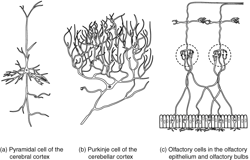
Neurons tin besides be classified on the ground of where they are establish, who establish them, what they do, or fifty-fifty what chemicals they utilise to communicate with each other. Some neurons referred to in this section on the nervous system are named on the basis of those sorts of classifications ((Figure)). For example, a multipolar neuron that has a very of import role to play in a part of the brain chosen the cerebellum is known as a Purkinje (commonly pronounced per-KIN-gee) cell. It is named after the anatomist who discovered it (January Evangilista Purkinje, 1787–1869).
Other Neuron Classifications
Three examples of neurons that are classified on the footing of other criteria. (a) The pyramidal cell is a multipolar cell with a jail cell torso that is shaped something similar a pyramid. (b) The Purkinje cell in the cerebellum was named after the scientist who originally described it. (c) Olfactory neurons are named for the functional grouping with which they belong.
Glial Cells
Glial cells, or neuroglia or simply glia, are the other type of cell found in nervous tissue. They are considered to exist supporting cells, and many functions are directed at helping neurons complete their function for communication. The proper name glia comes from the Greek word that means "mucilage," and was coined by the German language pathologist Rudolph Virchow, who wrote in 1856: "This connective substance, which is in the brain, the spinal cord, and the special sense nerves, is a kind of glue (neuroglia) in which the nervous elements are planted." Today, research into nervous tissue has shown that there are many deeper roles that these cells play. And research may find much more nearly them in the hereafter.
There are six types of glial cells. Four of them are plant in the CNS and two are institute in the PNS. (Effigy) outlines some common characteristics and functions.
| Glial Jail cell Types past Location and Basic Function | ||
|---|---|---|
| CNS glia | PNS glia | Bones role |
| Astrocyte | Satellite cell | Support |
| Oligodendrocyte | Schwann cell | Insulation, myelination |
| Microglia | – | Immune surveillance and phagocytosis |
| Ependymal cell | – | Creating CSF |
Glial Cells of the CNS
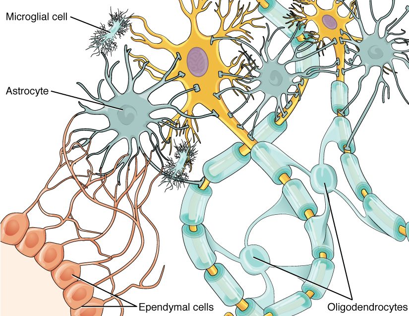
Ane jail cell providing back up to neurons of the CNS is the astrocyte, so named because it appears to be star-shaped under the microscope (astro- = "star"). Astrocytes have many processes extending from their main cell body (non axons or dendrites like neurons, but cell extensions). Those processes extend to interact with neurons, blood vessels, or the connective tissue covering the CNS that is called the pia mater ((Figure)). Generally, they are supporting cells for the neurons in the primal nervous system. Some ways in which they support neurons in the key nervous organization are by maintaining the concentration of chemicals in the extracellular infinite, removing excess signaling molecules, reacting to tissue damage, and contributing to the blood-brain barrier (BBB). The claret-brain barrier is a physiological barrier that keeps many substances that broadcast in the residual of the trunk from getting into the central nervous arrangement, restricting what can cantankerous from circulating blood into the CNS. Food molecules, such every bit glucose or amino acids, can pass through the BBB, but other molecules cannot. This really causes bug with drug delivery to the CNS. Pharmaceutical companies are challenged to design drugs that tin cross the BBB as well as have an effect on the nervous system.
Glial Cells of the CNS
The CNS has astrocytes, oligodendrocytes, microglia, and ependymal cells that back up the neurons of the CNS in several ways.
Like a few other parts of the body, the brain has a privileged claret supply. Very little tin pass through by diffusion. Most substances that cross the wall of a claret vessel into the CNS must exercise and so through an agile transport procedure. Because of this, only specific types of molecules tin enter the CNS. Glucose—the main free energy source—is allowed, as are amino acids. Water and another small particles, like gases and ions, can enter. But most everything else cannot, including white blood cells, which are i of the body'south main lines of defense. While this barrier protects the CNS from exposure to toxic or pathogenic substances, it besides keeps out the cells that could protect the brain and spinal cord from disease and damage. The BBB besides makes it harder for pharmaceuticals to be developed that tin can affect the nervous arrangement. Bated from finding efficacious substances, the ways of delivery is also crucial.
Besides found in CNS tissue is the oligodendrocyte, sometimes called just "oligo," which is the glial cell type that insulates axons in the CNS. The name means "cell of a few branches" (oligo- = "few"; dendro- = "branches"; -cyte = "jail cell"). At that place are a few processes that extend from the prison cell trunk. Each one reaches out and surrounds an axon to insulate it in myelin. One oligodendrocyte will provide the myelin for multiple axon segments, either for the same axon or for separate axons. The function of myelin volition exist discussed below.
Microglia are, equally the name implies, smaller than virtually of the other glial cells. Ongoing research into these cells, although not entirely conclusive, suggests that they may originate as white blood cells, called macrophages, that go office of the CNS during early on evolution. While their origin is not conclusively determined, their part is related to what macrophages practice in the rest of the torso. When macrophages encounter diseased or damaged cells in the residue of the body, they ingest and digest those cells or the pathogens that cause disease. Microglia are the cells in the CNS that can practise this in normal, healthy tissue, and they are therefore also referred to equally CNS-resident macrophages.
The ependymal cell is a glial cell that filters blood to make cerebrospinal fluid (CSF), the fluid that circulates through the CNS. Because of the privileged blood supply inherent in the BBB, the extracellular infinite in nervous tissue does non easily exchange components with the claret. Ependymal cells line each ventricle, one of four primal cavities that are remnants of the hollow middle of the neural tube formed during the embryonic development of the encephalon. The choroid plexus is a specialized construction in the ventricles where ependymal cells come up in contact with blood vessels and filter and absorb components of the blood to produce cerebrospinal fluid. Because of this, ependymal cells tin can exist considered a component of the BBB, or a place where the BBB breaks down. These glial cells announced similar to epithelial cells, making a single layer of cells with little intracellular space and tight connections between adjacent cells. They too have cilia on their apical surface to help move the CSF through the ventricular space. The relationship of these glial cells to the structure of the CNS is seen in (Effigy).
Glial Cells of the PNS
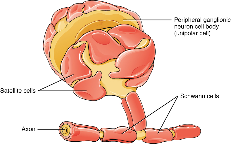
I of the two types of glial cells establish in the PNS is the satellite prison cell. Satellite cells are institute in sensory and autonomic ganglia, where they surround the cell bodies of neurons. This accounts for the name, based on their appearance under the microscope. They provide support, performing similar functions in the periphery as astrocytes do in the CNS—except, of grade, for establishing the BBB.
The second type of glial cell is the Schwann cell, which insulate axons with myelin in the periphery. Schwann cells are different than oligodendrocytes, in that a Schwann cell wraps effectually a portion of only 1 axon segment and no others. Oligodendrocytes have processes that reach out to multiple axon segments, whereas the entire Schwann cell surrounds just one axon segment. The nucleus and cytoplasm of the Schwann cell are on the edge of the myelin sheath. The relationship of these two types of glial cells to ganglia and nerves in the PNS is seen in (Effigy).
Glial Cells of the PNS
The PNS has satellite cells and Schwann cells.
Myelin
The insulation for axons in the nervous system is provided past glial cells, oligodendrocytes in the CNS, and Schwann cells in the PNS. Whereas the manner in which either cell is associated with the axon segment, or segments, that it insulates is different, the means of myelinating an axon segment is by and large the same in the two situations. Myelin is a lipid-rich sheath that surrounds the axon and by doing so creates a myelin sheath that facilitates the transmission of electrical signals along the axon. The lipids are essentially the phospholipids of the glial jail cell membrane. Myelin, nonetheless, is more than just the membrane of the glial prison cell. It also includes important proteins that are integral to that membrane. Some of the proteins help to hold the layers of the glial cell membrane closely together.
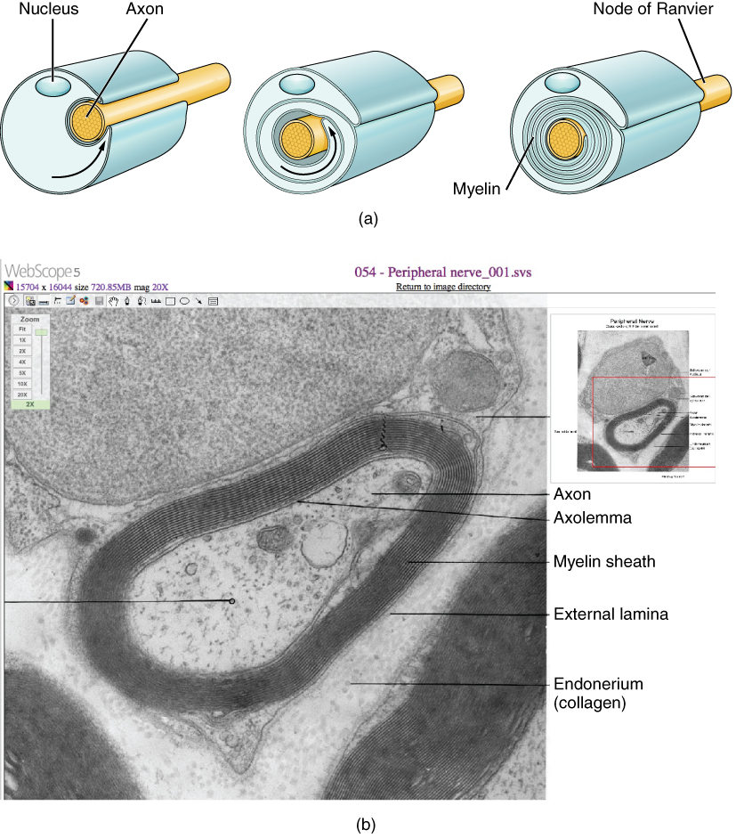
The advent of the myelin sheath tin can be thought of as similar to the pastry wrapped around a hot canis familiaris for "pigs in a blanket" or a similar nutrient. The glial cell is wrapped around the axon several times with little to no cytoplasm between the glial cell layers. For oligodendrocytes, the rest of the cell is separate from the myelin sheath every bit a cell process extends dorsum toward the cell body. A few other processes provide the same insulation for other axon segments in the area. For Schwann cells, the outermost layer of the cell membrane contains cytoplasm and the nucleus of the jail cell as a bulge on one side of the myelin sheath. During development, the glial cell is loosely or incompletely wrapped around the axon ((Figure)a). The edges of this loose enclosure extend toward each other, and one finish tucks under the other. The inner edge wraps effectually the axon, creating several layers, and the other edge closes effectually the outside and so that the axon is completely enclosed.
View the Academy of Michigan WebScope to see an electron micrograph of a cross-section of a myelinated nerve fiber. The axon contains microtubules and neurofilaments that are bounded by a plasma membrane known as the axolemma. Outside the plasma membrane of the axon is the myelin sheath, which is composed of the tightly wrapped plasma membrane of a Schwann cell. What aspects of the cells in this image react with the stain to brand them a deep, nighttime, black colour, such as the multiple layers that are the myelin sheath?
Myelin sheaths can extend for one or two millimeters, depending on the bore of the axon. Axon diameters can be as pocket-sized as 1 to 20 micrometers. Considering a micrometer is 1/g of a millimeter, this means that the length of a myelin sheath can be 100–1000 times the diameter of the axon. (Effigy), (Effigy), and (Effigy) evidence the myelin sheath surrounding an axon segment, merely are not to scale. If the myelin sheath were drawn to scale, the neuron would have to be immense—peradventure covering an entire wall of the room in which you are sitting.
The Process of Myelination
Myelinating glia wrap several layers of cell membrane around the cell membrane of an axon segment. A unmarried Schwann cell insulates a segment of a peripheral nerve, whereas in the CNS, an oligodendrocyte may provide insulation for a few separate axon segments. EM × 1,460,000. (Micrograph provided by the Regents of Academy of Michigan Medical School © 2012)
Disorders of the…
Nervous Tissue Several diseases tin outcome from the demyelination of axons. The causes of these diseases are not the same; some have genetic causes, some are caused by pathogens, and others are the result of autoimmune disorders. Though the causes are varied, the results are largely similar. The myelin insulation of axons is compromised, making electrical signaling slower.
Multiple sclerosis (MS) is one such disease. It is an example of an autoimmune disease. The antibodies produced by lymphocytes (a blazon of white claret cell) marking myelin as something that should not be in the body. This causes inflammation and the destruction of the myelin in the central nervous system. Every bit the insulation around the axons is destroyed by the disease, scarring becomes obvious. This is where the name of the disease comes from; sclerosis means hardening of tissue, which is what a scar is. Multiple scars are establish in the white matter of the encephalon and spinal string. The symptoms of MS include both somatic and autonomic deficits. Control of the musculature is compromised, as is control of organs such as the float.
Guillain-Barré (pronounced gee-YAN bah-RAY) syndrome is an instance of a demyelinating affliction of the peripheral nervous arrangement. It is as well the result of an autoimmune reaction, simply the inflammation is in peripheral nerves. Sensory symptoms or motor deficits are common, and autonomic failures tin can pb to changes in the heart rhythm or a driblet in blood force per unit area, especially when standing, which causes dizziness.
Chapter Review
Nervous tissue contains two major jail cell types, neurons and glial cells. Neurons are the cells responsible for communication through electrical signals. Glial cells are supporting cells, maintaining the surround around the neurons.
Neurons are polarized cells, based on the flow of electrical signals forth their membrane. Signals are received at the dendrites, are passed along the cell torso, and propagate along the axon towards the target, which may be another neuron, muscle tissue, or a gland. Many axons are insulated by a lipid-rich substance chosen myelin. Specific types of glial cells provide this insulation.
Several types of glial cells are institute in the nervous system, and they can exist categorized past the anatomical segmentation in which they are found. In the CNS, astrocytes, oligodendrocytes, microglia, and ependymal cells are plant. Astrocytes are important for maintaining the chemic environment effectually the neuron and are crucial for regulating the blood-brain barrier. Oligodendrocytes are the myelinating glia in the CNS. Microglia act every bit phagocytes and play a part in immune surveillance. Ependymal cells are responsible for filtering the blood to produce cerebrospinal fluid, which is a circulatory fluid that performs some of the functions of blood in the brain and spinal string considering of the BBB. In the PNS, satellite cells are supporting cells for the neurons, and Schwann cells insulate peripheral axons.
Interactive Link Questions
Visit this site to larn almost how nervous tissue is composed of neurons and glial cells. The neurons are dynamic cells with the ability to make a vast number of connections and to respond incredibly quickly to stimuli and to initiate movements based on those stimuli. They are the focus of intense inquiry as failures in physiology tin atomic number 82 to devastating illnesses. Why are neurons only constitute in animals? Based on what this article says about neuron function, why wouldn't they be helpful for plants or microorganisms?
Neurons enable thought, perception, and move. Plants exercise not move, and then they practise not need this blazon of tissue. Microorganisms are likewise small to have a nervous arrangement. Many are single-celled, and therefore have organelles for perception and movement.
View the University of Michigan Webscope to see an electron micrograph of a cross-section of a myelinated nerve cobweb. The axon contains microtubules and neurofilaments, bounded past a plasma membrane known as the axolemma. Exterior the plasma membrane of the axon is the myelin sheath, which is composed of the tightly wrapped plasma membrane of a Schwann jail cell. What aspects of the cells in this image react with the stain that makes them the deep, dark, black color, such as the multiple layers that are the myelin sheath?
Lipid membranes, such every bit the cell membrane and organelle membranes.
Review Questions
What type of glial jail cell provides myelin for the axons in a tract?
- oligodendrocyte
- astrocyte
- Schwann prison cell
- satellite cell
Which part of a neuron contains the nucleus?
- dendrite
- soma
- axon
- synaptic end bulb
Which of the following substances is least able to cross the blood-brain barrier?
- water
- sodium ions
- glucose
- white blood cells
What blazon of glial cell is the resident macrophage behind the claret-encephalon barrier?
- microglia
- astrocyte
- Schwann cell
- satellite cell
What two types of macromolecules are the main components of myelin?
- carbohydrates and lipids
- proteins and nucleic acids
- lipids and proteins
- carbohydrates and nucleic acids
Critical Thinking Questions
Multiple sclerosis is a demyelinating disease affecting the key nervous system. What type of cell would exist the almost likely target of this disease? Why?
The disease would target oligodendrocytes. In the CNS, oligodendrocytes provide the myelin for axons.
Which type of neuron, based on its shape, is best suited for relaying information directly from one neuron to another? Explain why.
Bipolar cells, because they have one dendrite that receives input and one axon that provides output, would be a direct relay between two other cells.
Glossary
- astrocyte
- glial cell type of the CNS that provides support for neurons and maintains the blood-brain bulwark
- axon hillock
- tapering of the neuron prison cell body that gives rise to the axon
- axon segment
- single stretch of the axon insulated by myelin and bounded by nodes of Ranvier at either stop (except for the first, which is subsequently the initial segment, and the final, which is followed past the axon terminal)
- axon final
- stop of the axon, where there are usually several branches extending toward the target cell
- axoplasm
- cytoplasm of an axon, which is different in composition than the cytoplasm of the neuronal cell body
- bipolar
- shape of a neuron with two processes extending from the neuron prison cell body—the axon and ane dendrite
- blood-brain barrier (BBB)
- physiological bulwark between the circulatory system and the central nervous system that establishes a privileged claret supply, restricting the flow of substances into the CNS
- cerebrospinal fluid (CSF)
- circulatory medium within the CNS that is produced past ependymal cells in the choroid plexus filtering the claret
- choroid plexus
- specialized structure containing ependymal cells that line blood capillaries and filter blood to produce CSF in the four ventricles of the brain
- ependymal cell
- glial cell blazon in the CNS responsible for producing cerebrospinal fluid
- initial segment
- first part of the axon as it emerges from the axon hillock, where the electrical signals known as action potentials are generated
- microglia
- glial jail cell type in the CNS that serves as the resident component of the allowed system
- multipolar
- shape of a neuron that has multiple processes—the axon and two or more dendrites
- myelin sheath
- lipid-rich layer of insulation that surrounds an axon, formed by oligodendrocytes in the CNS and Schwann cells in the PNS; facilitates the transmission of electrical signals
- node of Ranvier
- gap between two myelinated regions of an axon, allowing for strengthening of the electrical betoken equally it propagates down the axon
- oligodendrocyte
- glial cell type in the CNS that provides the myelin insulation for axons in tracts
- satellite prison cell
- glial prison cell type in the PNS that provides support for neurons in the ganglia
- Schwann cell
- glial prison cell type in the PNS that provides the myelin insulation for axons in fretfulness
- synapse
- narrow junction across which a chemic indicate passes from neuron to the next, initiating a new electrical bespeak in the target cell
- synaptic end bulb
- swelling at the end of an axon where neurotransmitter molecules are released onto a target cell across a synapse
- unipolar
- shape of a neuron which has only one process that includes both the axon and dendrite
- ventricle
- cardinal cavity inside the brain where CSF is produced and circulates
lambertsequal1961.blogspot.com
Source: https://opentextbc.ca/anatomyandphysiologyopenstax/chapter/nervous-tissue/
0 Response to "Histology of Nervous Tissue Review Sheet Answer Key"
Post a Comment